Automatic Classification of Artery/Vein from Single Wavelength Fundus Images
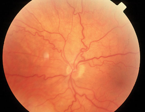
Vessels are regions of prominent interest in retinal fundus images. Classification of vessels into arteries and veins can be used to assess the oxygen saturation level, which is one of the indicators for the risk of stroke, condition of diabetic retinopathy, and hypertension. In practice, dual-wavelength images are obtained to emphasize arteries and veins separately. In this paper, we propose an automated technique for the classification of arteries and veins from single-wavelength fundus images using convolutional neural networks employing the ResNet-50 backbone and squeeze-excite blocks. We formulate the artery-vein identification problem as a three-class classification problem where each pixel is labeled as belonging to an artery, vein, or the background. The proposed method is trained on publicly available fundus image datasets, namely RITE, LES-AV, IOSTAR, and cross-validated on the HRF dataset. The standard performance metrics, such as average sensitivity, specificity, accuracy, and area under the curve for the datasets mentioned above, are 92.8%, 93.4%, 93.4%, and 97.5%, respectively, which are superior to the state-of-the-art methods.
Published in: 2020 IEEE 17th International Symposium on Biomedical Imaging (ISBI)
Date of Conference: 3-7 April 2020
Date Added to IEEE Xplore: 22 May 2020
NSPEC Accession Number: 19725889
DOI: 10.1109/ISBI45749.2020.9098580
Publisher: IEEEConference Location: Iowa City, IA, USA