Infyuva Tech Solutions
Excellence in Healthcare AI Solutions
About
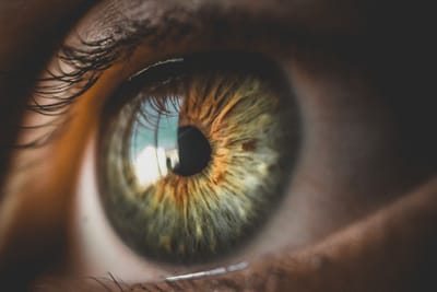
We, at Infyuva, developing a complete iCare smartphone-based hardware and software solution that can cater to a wide range of retinal pathologies such as glaucoma, diabetic retinopathy, macular degeneration, micro aneurysm detection, retinal oxygen saturation estimation, and many more.
our solutions
iCare: Smart-Phone based Solutions
product Deck
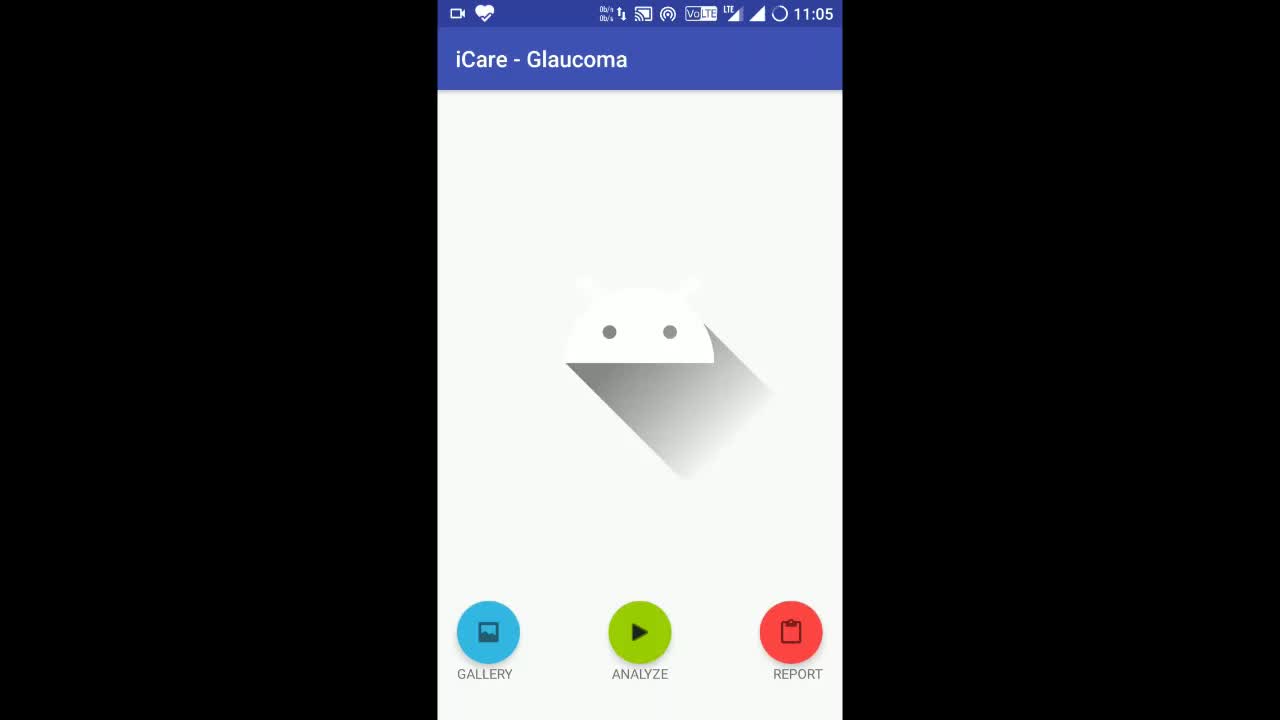
GLAUCOMA PRESCREEING APP
Events
Releasing Soon!!!
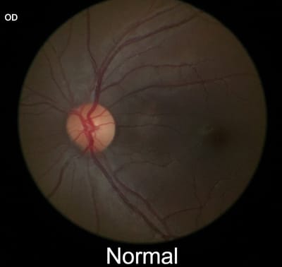
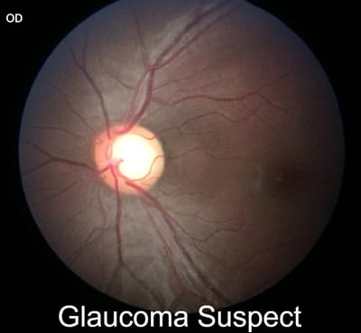
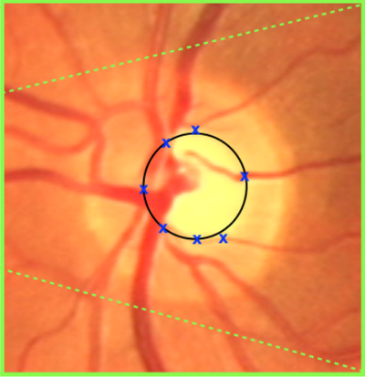
Retinal fundus images are critical in diagnosis of vision-related diseases such as glaucoma, diabetic retinopathy, macular degeneration, macular edema, diabetic maculopathy, etc. Retinal fundus examination also makes it to predict neurological and cardiovascular disorders. Although several fundus image databases are available, there is still a lack of glaucoma-specific fundus image database with a large number of images having manual annotations (ground-truth) of both optic disc (OD) and optic cup (OC) and also a decision on the glaucoma severity. We have created a comprehensive and large glaucoma-specific retinal fundus images database with 1500 images corresponding to both glaucomatous and normal conditions. We provide OD and OC manual segmentation ground-truth and a decision on glaucoma severity grading by five expert ophthalmologists. The database will be made publicly available very soon to enable fellow researchers to validate their segmentation and classification algorithms.
Articles
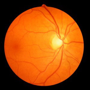
A novel and fully automated fundus image processing technique for glaucoma prescreening based on the rim-to-disc ratio (RDR).
Read More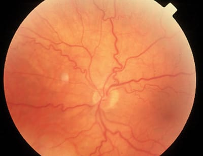
Classification of vessels into arteries and veins can be used to assess the oxygen saturation level, condition of diabetic retinopathy, and hypertension
Read More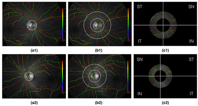
An automatic technique for the measurement of oxygen saturation in retinal arterioles and venules using dual-wavelength retinal oximetry images.
Read More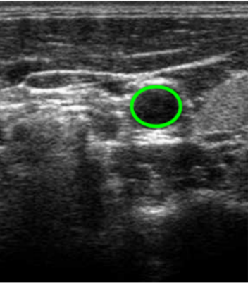
Elliptical active disc technique for the segmentation of common carotid artery lumen intima layer from transverse mode ultrasound images.
Read More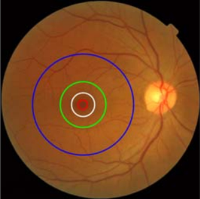
Contact
- Infyuva Tech Solutions Pvt. Ltd, MUTBI, 4TH FLOOR, NO. 21/1, ADVANCED RESEARCH CENTER, MADHAVA NAGAR, MANIPAL, UDUPI, KARNATAKA, INDIA
- 9945575205
- 9902762074
- infyuva@gmail.com
- 9 - 6 pm