Macular Degeneration
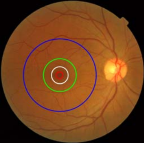
The macula is a central area in the retina of the human eye. The center of the macula is called the fovea and is responsible for central, high-resolution vision. The macula is subdivided into regions called foveal avascular zone (FAZ), fovea, parafovea, and perifovea. The foveal avascular zone can further be subdivided into umbo and foveola regions. Figure 1 shows the retinal fundus image, with the different regions of the macula outlined. These regions are described and delineated on the basis of histological findings. They are hard to differentiate using a fundus image of the retina. Certain pathological conditions of the macula such as age-related macular degeneration (AMD), diabetic macular edema, macular hole, and macular pucker can affect the macula and, in turn, one’s central vision.
Maculopathy is one of the leading causes of vision loss]. For identification of the stage and severity of maculopathy, it is important to segment the fovea and its surrounding regions within the macula. Fundus imaging machines are an effective tool for diagnosing retinal diseases such as maculopathy, but there is a lack of robust, semi-automated or automated disease diagnosis tools. Some efforts have been made to develop an automated or semi-automated technique for reliable segmentation of fovea followed by macular region delineation and subsequent diagnosis of the stage and severity of maculopathy.
We have developed a fully automated technique for segmentation and delineation of macular regions using retinal fundus images, which is a precursor and an important step in the severity grading of maculopathy.